World Stroke Day 2026: All About AVMs And Brain Haemorrhage
Total Views |


Brain arteriovenous malformations (AVMs) are developmental abnormalities in which arteries connect directly to veins, bypassing the normal network of tiny blood vessels called capillaries that supply oxygen and nutrients to brain tissue. This tangle of abnormal vessels, known as the nidus, can remain undetected for years but may sometimes rupture, causing a potentially life-threatening brain haemorrhage.
What Are The Symptoms?
Warning signs include a sudden, severe headache, seizures, loss of consciousness or new neurological problems, all of which require immediate medical attention. Some AVMs may also cause vision disturbances or tinnitus, while others are discovered incidentally.
“Brain AVMs may initially start small in infancy and early childhood and grow with the child’s development, as well as due to the abnormally high blood flow within the AVM,” said Dr. Chinmay Nagesh, Senior Consultant in Stroke and Neurovascular Intervention and Endovascular Interventional Radiology at Apollo Speciality Hospital, Bengaluru.
Accurate imaging and timely specialist evaluation help doctors assess the AVM and decide whether treatment is needed and which approach is most suitable, including embolisation in selected cases. As not every AVM requires the same approach, management must be individualised, balancing the risks posed by the malformation against those of intervention. Regular follow-up helps doctors monitor the patient’s condition and check the treatment results.
What To Do When An AVM Ruptures
“When an AVM ruptures and causes a brain haemorrhage, the immediate priority is to stabilise the patient,” said Dr Vikram Huded, President of the Indian Stroke Association; Senior Consultant, Director and Clinical Lead of the Interventional Neurology Programme at Narayana Health Group; Governing Council Lead of Neuroscience; and Head of Neurology, Bengaluru.
Treatment may involve securing the airway and circulation, controlling blood pressure and preventing further brain injury. Doctors also manage complications such as raised pressure inside the skull, seizures and hydrocephalus, a build-up of fluid in the brain. Some patients may require emergency surgery or other procedures to relieve pressure or control bleeding. Urgent evaluation by a multidisciplinary team of stroke specialists, neurosurgeons and neurointerventional specialists is essential.
Why Are Some Brain AVMs Difficult To Treat?
Brain AVMs vary in complexity. Those located deep within the brain or near areas controlling speech, movement or vision are particularly challenging because treatment-related injury can cause significant disability. Dr. Chinmay said large or diffused AVMs, those supplied by multiple small or tortuous arteries, and those with complex deep venous drainage may be difficult to treat safely through embolisation alone. Associated aneurysms and fragile blood vessels can further complicate treatment.
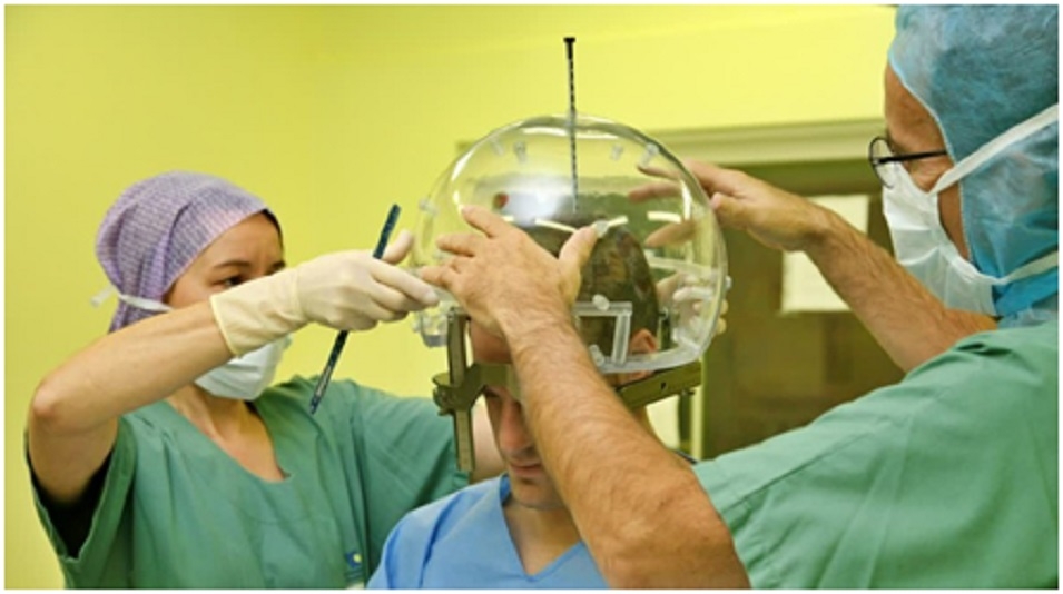
Experts say treatment choice depends on the AVM's size, location, and structure; the patient's age and overall health; history of haemorrhage; and the risks associated with each treatment option. Dr. Chinmay and Dr Huded explain that microsurgery can immediately remove an AVM if it is surgically accessible. Endovascular embolisation uses a catheter to block abnormal vessels and may be performed alone or in combination with surgery or radiosurgery. Embolisation alone may be suitable when complete and safe closure is achievable. Stereotactic radiosurgery uses focused radiation to close abnormal vessels gradually and may be considered for smaller AVMs or those in difficult-to-reach locations. As this process takes time, the risk of haemorrhage may persist until the AVM is completely obliterated.